| << |
1 |
2 |
3 |
4 |
5 |
6 |
7 |
8 |
9 |
10 |
11 |
12 |
13 |
14 |
15 |
16 |
17 |
18 |
19 |
20 |
21 |
22 |
23 |
24 |
25 |
26 |
27 |
28 |
29 |
30 |
31 |
Deep-Sea Bugs! 2/1/2006
By David Pearce
The microbial community is primarily responsible
for recycling the nutrients which reach the sea floor. For
this reason, a group of three scientists from the British
Antarctic Survey (BAS) are investigating the biodiversity
and community structure of microorganisms within the sediment
and comparing the nutrient rich (M5) and nutrient poor (M6)
sites around the Crozet Islands. This work will involve microscopy
during the cruise itself, with stains that selectively mark
key groups of bacteria and this will be complimented with
a range of DNA based molecular techniques back in the laboratories
at BAS.
|
|
 |
|
|
Robin
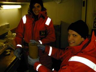
and Rachel demonstrate the delicate art of slicing mud!
Click on the pictures for a larger version |
Bacteria are very small (< 100th of a millimetre
in length) and so it is often impossible to describe their
taxonomy using morphological (or cell shape) characteristics
alone. For this reason, we can design specific chemicals (oligonucleotide
probes) which bind to particular DNA sequences which only
occur in selected groups of bacteria. If these chemicals are
synthesised to include a dye which fluoresces (glows) under
ultra-violet light, then only those cells which contain a
specific DNA sequence are stained by that chemical. These
can then be detected using a special type of microscope with
appropriate filters. We will also be using two further fluorescent
stains: DAPI to stain all DNA in the sample, and hence estimate
the total population density, and CTC which will fluoresce
only in living cells, to estimate how many cells have survived
the ascent to the surface. In this way we can start to build
up a picture of the community structure whilst still at sea.
On return to the laboratories in Cambridge,
we will extract the total community DNA from the sediment.
This will be used to identify the dominant groups of bacteria
present within the sample (using denaturing gradient gel electrophoresis
– a technique which requires about 5000 cells per ml
before identification can take place) and to construct clone
libraries, which, with the assistance of a little statistics,
can be used to estimate the total microbial biodiversity across
each site.
 |