University
of LiverpoolUniversity
of Liverpool
University
of LiverpoolUniversity
of Liverpool[ Dissection Home | Fishweb]
(This is also the Starting Point for the Zoology Honours Display Prac Virtual
Dissection
2002
for students who do not wish to do 'real' dissection)
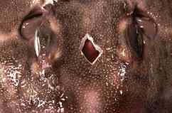
| A diamond shaped hole should be cut between the eyes at the end of the first practical to enable penetration of fixative |
 |
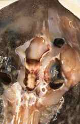
| The first stage is to remove the skin over the cranial area |
|
| The brain is exposed by shaving away the cranial cartilage. Shave away the otic capsule cutting down through the semi-circular canals |
|
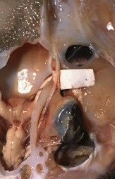
| The nerves can be seen through the cartilage here the superficial opthalmic has been as it crosses over the opthalmic tract (various cranial
nerves can be seen plus the |
|
| After removal of the eye, the nerves can be seen crossing the floor of the orbit (note the otolith exposed within the auditory capsule) |
|
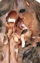
| Close-up view of the floor of the orbit and brain |
|
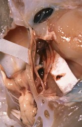
| The dissection has been extended by shaving away the spinal column revealing the spinal nerves. The vagus has also been exposed. |
|