School of Environmental Sciences
 SEM images
 Scanning Electron Microscope (SEM)
The SEM is a type of electron microscope that images a sample by scanning it with a very narrow beam of electrons.
This type of microscope uses a focused beam of high-energy electrons which generates a range of signals when it hits
a solid object and interacts with its surface. These signals provide information about the texture and chemical
composition of the sample as well as details of the crystalline structure and orientation of the materials in the sample.
Areas ranging from approximately 1 cm to 5 microns in width can be imaged by the SEM with magnification ranging
from 20X to 30,000X. The SEM may produce very high-resolution images of a sample, showing details less than 1 nm
in size and, because SEM images have a large depth of field, these images indicate the three-dimensional appearance
of the sample’s surface.
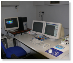
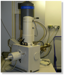
Photographs here show the
Philips XL 30 SEM with a four
element KE backscatter detector
and a link ISIS x-ray diffraction
detector.
The other advantages of using
the scanning electron microscope
are that it also allows us to:
•
analyse selected point
locations on the sample,
•
show spatial variations in
chemical compositions and
•
produce elemental maps
for the sample.
.
.
Scanning Electron Microscope (SEM)
The SEM is a type of electron microscope that images a sample by scanning it with a very narrow beam of electrons.
This type of microscope uses a focused beam of high-energy electrons which generates a range of signals when it hits
a solid object and interacts with its surface. These signals provide information about the texture and chemical
composition of the sample as well as details of the crystalline structure and orientation of the materials in the sample.
Areas ranging from approximately 1 cm to 5 microns in width can be imaged by the SEM with magnification ranging
from 20X to 30,000X. The SEM may produce very high-resolution images of a sample, showing details less than 1 nm
in size and, because SEM images have a large depth of field, these images indicate the three-dimensional appearance
of the sample’s surface.
Photographs here show the
Philips XL 30 SEM with a four
element KE backscatter detector
and a link ISIS x-ray diffraction
detector.
The other advantages of using
the scanning electron microscope
are that it also allows us to:
•
analyse selected point
locations on the sample,
•
show spatial variations in
chemical compositions and
•
produce elemental maps
for the sample.
.
.
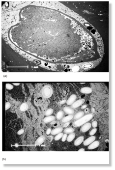
 SEM images
The SEM images (a) and (b) were
taken on a thin section of the
Jurassic Osmington Miils
Ironstone Formation, Dorset.
The third photograph is taken on
a sample of the materials found in
one of the research tanks at the
University of Liverpool’s Botanical
Gardens at Ness, South Wirral.
Here is a link to a website showing the images taken by Alex Cairns during
his work experience placement in the Department of Earth and Ocean
Sciences.
If you would like more information about how you to arrange a work
experience placement in Earth and Ocean Sciences, please contact Chris
Hunt in the School of Environmental Sciences by sending an email to:
scfc@liverpool.ac.uk
SEM images
The SEM images (a) and (b) were
taken on a thin section of the
Jurassic Osmington Miils
Ironstone Formation, Dorset.
The third photograph is taken on
a sample of the materials found in
one of the research tanks at the
University of Liverpool’s Botanical
Gardens at Ness, South Wirral.
Here is a link to a website showing the images taken by Alex Cairns during
his work experience placement in the Department of Earth and Ocean
Sciences.
If you would like more information about how you to arrange a work
experience placement in Earth and Ocean Sciences, please contact Chris
Hunt in the School of Environmental Sciences by sending an email to:
scfc@liverpool.ac.uk