Eosinophilic enteritis may occur as part of a more generalised disease process
where a heavy infiltration of eosinophils are seen in multiple organs e.g. skin
as well as variable portions of the gastro-intestinal tract. Eosinophilic infiltration
may affect solely the gastro-intestinal tract in a diffuse fashion (usually
manifesting as a malassimilation or malabsorption syndrome) or at a more focal
site e.g. segmental lesions of the large intestine or small intestine.
Focal eosinophilic enteritis lesions have been associated with nematode larvae and Pythium spp. but several cases in the literature have had no obvious inciting cause.
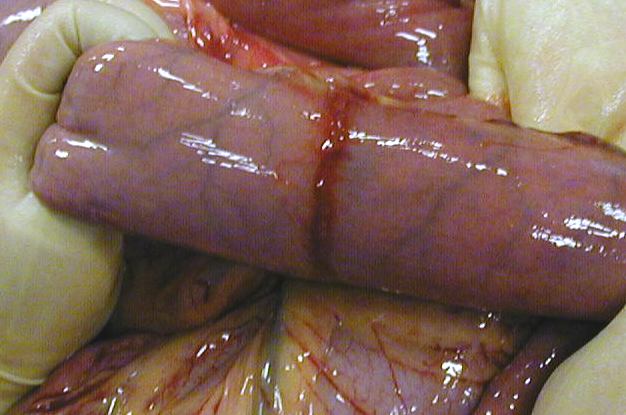
These idiopathic, focal eosinophilic enteritis lesions usually present with colic. Ingesta becomes obstructed at the site which may involve any region of the small intestine. The lesions can be circumferential or plaque like in nature and may be multiple. Few are reported in the literature but at this clinic we have seen an increasing number over the past 3 years.
|
This is an example of a circumferential eosinophilic enteritis lesion
Relevant references :
Southwood L.L., Kawcak C.E., Trotter G.W., Stashak T.S. and Frisbee D.D. (2000)
Idiopathic focal eosinophilic enteritis associated with small intestinal obstruction
in 6 horses. Vet. Surg. 29, 415-419
Stanar L.S., Little D., Redding W.R. and Jones S.L. (2002) Idiopathic eosinophilic enteritis in a 10 week old colt. Compend. Contin. Educ. Pract. Vet, 342-344
Swain J.M., Licka T., Rhind S.M. and Hudson N.P.H. (2003) Multifocal eosinophilic enteritis associated with a small intestinal obstruction in a standardbred horse. Vet. Rec.152, 648-651